Abstract
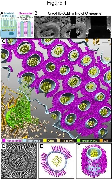
Microvilli are actin-bundle-supported membrane protrusions essential for absorption, secretion, and sensation. Microvilli defects cause gastrointestinal disorders; however, mechanisms controlling microvilli formation and organization remain unresolved. Here, we study microvilli by vitrifying the Caenorhabditis elegans larvae and mouse intestinal tissues with high-pressure freezing, thinning them with cryo-focused ion-beam milling, followed by cryo-electron tomography and subtomogram averaging. We find that many radial nanometer bristles referred to as nanobristles project from the lateral surface of nematode and mouse microvilli. The C. elegans nanobristles are 37.5 nm long and 4.5 nm wide. Nanobristle formation requires a protocadherin family protein, CDH-8, in C. elegans. The loss of nanobristles in cdh-8 mutants slows down animal growth and ectopically increases the number of Y-shaped microvilli, the putative intermediate structures if microvilli split from tips. Our results reveal a potential role of nanobristles in separating microvilli and suggest that microvilli division may help generate nascent microvilli with uniformity.
|
Paper Link:https://www.pnas.org/doi/10.1073/pnas.2122249119
|