In the cerebral cortex, sensory processing of external stimuli can evoke neural networks to fire flurried electrical responses. These responses can leave neurological traces from a particular experience like crumb trails, which can be strengthened for long-term recall. However, due to the complexity of the connections and sheer number of neurons within the cortex, understanding these traces has been daunting for neuroscientists.
In a new study, researchers have combined 3D imaging with computational methods to simultaneously track how thousands of neurons respond to external stimuli in the cortex of mice, long-term. For the first time, the study showed long-term memory traces formed and reactivated by a small portion of neurons in the cortex’s second layer. That team later reported activation to be sparsely distributed across many cortical areas.
The findings are published in Proceedings of the National Academy of Sciences.
“There are a lot of activities going on in the cortex and it’s really hard to piece out which neurons are responsible for long-term memory,” said the study’s corresponding author, JiSong Guan, Principle Investigator at the Center for Life Sciences at Tsinghua University in China. “For the first time, we’ve directly shown how context environments can specifically activate a very small portion of neurons, and this can be reactivated during recall of memory.”
Guan says that of the six cortical layers, layer two has been considered a ‘high-function’ layer, responsible for certain information processing. However, its specific function has remained unknown.
“We haven’t known where memory is stored so that has been a black box, but now with this method we are able to look at the neuronal circuit to see what is responsible for this cognitive function,” said Guan.
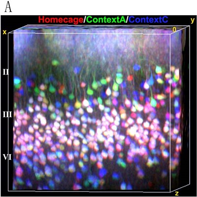
In their study, the team first began familiarizing mice with three distinct environment-specific trials over a two-month period. To track the neurons activated in cortical circuits during each behavioral trial, the team used florescent tagging of early growth response protein, EGR1 – a protein normally expressed during high-frequency neuronal stimulation and long-term learning processes.
By highlighting the expression of EGR1, the lab used two-photon imaging technology to visualize the neuronal activity in 3D slices across single cortical volumes. Then computationally, Guan’s team utilized a newly developed automatic recognition algorithm to quantitate and reconstruct the activity of thousands of individual neurons in each mouse over the long-term.
The team ultimately found task-specific neuronal activation in cortical layer II, but was surprised to see later activation in multiple cortical regions.
“We were originally expecting some specific region of the cortex to have a strong response of a typical memory because some areas are more important than others,” explained Guan. “But we found that context memories show specific storage in almost each individual cortical area as a sparse response, so that was kind of surprising to us.”
Guan says their technology could be applied to study diseases like autism or schizophrenia by observing how neural networks responses change in mouse models with physiological disease. For now, Guan’s lab will investigate how cortical layer II is forming memory traces.
“They must come from other layers and we want to better understand how those neural circuits gather together to form a specific response to a very complicated environment,” said Guan. ?“How those memories change their location and gradually build the remote memory in the cortical is still an important question to ask.”